Key Takeaways:

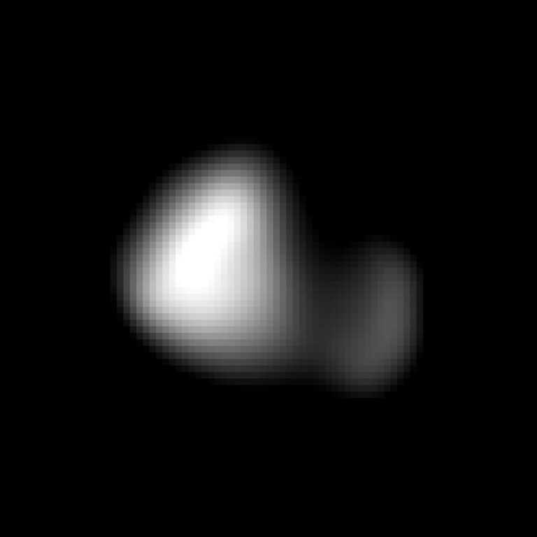
NASA’s Phoenix Mars Lander has taken the first-ever image of a single particle of Mars’ ubiquitous dust, using its atomic force microscope.
The particle — shown at higher magnification than anything ever seen from another world — is a rounded particle about one micrometer, or one millionth of a meter, across. It is a speck of the dust that cloaks Mars. Such dust particles color the martian sky pink, feed storms that regularly envelop the planet and produce Mars’ distinctive red soil.
“This is the first picture of a clay-sized particle on Mars, and the size agrees with predictions from the colors seen in sunsets on the Red Planet,” says Phoenix coinvestigator Urs Staufer of the University of Neuchatel, Switzerland, who leads a Swiss consortium that made the microscope.
“Taking this image required the highest resolution microscope operated off Earth and a specially designed substrate to hold the Martian dust,” says Tom Pike, Phoenix science team member from Imperial College London. “We always knew it was going to be technically very challenging to image particles this small.”
It took a very long time, roughly a dozen years, to develop the device that is operating in a polar region on a planet now about 220 million miles away (350 million kilometers).
The atomic force microscope maps the shape of particles in three dimensions by scanning them with a sharp tip at the end of a spring. During the scan, invisibly fine particles are held by a series of pits etched into a substrate microfabricated from a silicon wafer. Pike’s group at Imperial College produced these silicon microdiscs.
The atomic force microscope can detail the shapes of particles as small as about 100 nanometers, about one one-thousandth the width of a human hair. That is about 100 times greater magnification than seen with Phoenix’s optical microscope, which made its first images of Martian soil about 2 months ago. Until now, Phoenix’s optical microscope held the record for producing the most highly magnified images to come from another planet.
“I’m delighted that this microscope is producing images that will help us understand Mars at the highest detail ever,” Staufer says. “This is proof of the microscope’s potential. We are now ready to start doing scientific experiments that will add a new dimension to measurements being made by other Phoenix lander instruments.”
“After this first success, we’re now working on building up a portrait gallery of the dust on Mars,” Pike adds.
Mars’ ultra-fine dust is the medium that actively links gases in the martian atmosphere to processes in martian soil, so it is critically important to understanding Mars’ environment, the researchers say.
The particle seen in the atomic force microscope image was part of a sample scooped by the robotic arm from the “Snow White” trench and delivered to Phoenix’s microscope station in early July. The microscope station includes the optical microscope, the atomic force microscope and the sample delivery wheel. It is part of a suite of tools called Phoenix’s Microscopy, Electrochemistry and Conductivity Analyzer.